Anatomical description of Peyer's patches in the small intestine of picuro (Cuniculus paca)
DOI:
https://doi.org/10.55873/ariva.v2i2.251Keywords:
anatomy Peyer’s patches, picuroAbstract
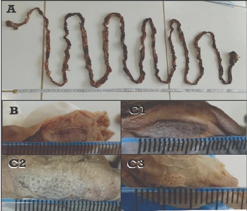
In order to describe anatomical aspects of Peyer's patches in picuro (Cuniculus paca), 10 samples of small intestine of picuros from rural communities in the regionof Tambopata -Madre de Dios were collected and analyzed. The results showed that the Peyer's patches in picuros are isolated and independent structures that are distributed throughout the small intestine, with respect to the transverse plane of the intestine, mainly in the antimesenteric border and randomly with respect to the longitudinal plane; Although the number and size of Peyer's patches show variability, the best records are observed at the level of the jejunum, with a significant difference (p<0.05) in relation to the other segments. Concluding that the Peyer's patches of picuros(Cuniculus paca)are anatomically peculiar among many mammals, but they resemble animals classified in group II such as rodents, non-human primates and rabbits.
References
Aquino, R., Gil, D., & Pezo, E. (2009). Aspectos ecológicos y sostenibilidad de la caza del majás (Cuniculus paca) en la cuenca del río Itaya, amazonía peruana. Revista Peruana de Biología, 16(1).
Brandtzaeg, P., Kiyono, H., Pabst, R., & Russell, M. . (2008). Terminology: nomenclature of mucosa-associated lymphoid tissue. Mucosal Immunology, 1(1), 31-37. https://doi.org/10.1038/mi.2007.9
Cesta, M. F. (2006). Normal structure, function, and histology of mucosa-associated Lymphoid tissue. Toxicologic Pathology, 34(5), 599-608. https://doi.org/10.1080/01926230600865531
Flores, J., Navarrete, M., & Sato, A. (2020). Descripción anatómica de las placas de Peyer en el intestino delgado de la alpaca (Vicugna pacos). Revista de Investigaciones Veterinarias del Perú, 31(3), e18175. https://doi.org/10.15381/rivep.v31i3.18175
Gebert, A., Rothkötter, H.-J., & Pabst, R. (1996). M Cells in Peyer’s Patches of the Intestine. En Kwang W. Jeon (Ed.), International Review of Cytology (pp. 91-159). Academic Press. https://doi.org/10.1016/S0074-7696(08)61346-7
Getty, R. (2001). Anatomía de los animales domésticos (5ta ed.). Masson S.A.
Gil, J., Gimeno, M., Laborda, J., & Nuviala, J. (2012). Protocolos de disección (3era ed.). SERVET.
Haley, P. J. (2017). The lymphoid system: a review of species differences. Journal of Toxicologic Pathology, 30(2), 111-123. https://doi.org/10.1293/tox.2016-0075
Heel, K. A., Mccauley, R. ., Papadimitriou, J. ., & Hall, J. . (1997). REVIEW: Peyer’s patches. Journal of Gastroenterology and Hepatology, 12(2), 122-136. https://doi.org/10.1111/j.1440-1746.1997.tb00395.x
International Committee on Veterinary Gross Anatomical Nomenclature (ICVGAN). (2017). Nomina anatomica veterinaria (6ta ed.). World Association of Veterinary Anatomists.
Jung, C., Hugot, J.-P., & Barreau, F. (2010). Peyer’s Patches: The Immune Sensors of the Intestine. International Journal of Inflammation, 2010, 1-12. https://doi.org/10.4061/2010/823710
Landsverk, T., Halleraker, M., Aleksandersen, M., McClure, S., Hein, W., & Nicander, L. (1991). The intestinal habitat for organized lymphoid tissues in ruminants; comparative aspects of structure, function and development. Veterinary Immunology and Immunopathology, 28(1), 1-16. https://doi.org/10.1016/0165-2427(91)90038-E
Liebler-Tenorio, E. M., & Pabst, R. (2006). MALT structure and function in farm animals. Veterinary Research, 37(3), 257-280. https://doi.org/10.1051/vetres:2006001
Michalski, F., & Norris, D. (2011). Activity pattern of Cuniculus paca (Rodentia: Cuniculidae) in relation to lunar illumination and other abiotic variables in the southern Brazilian Amazon. Zoologia (Curitiba), 28(6), 701-708. https://doi.org/10.1590/S1984-46702011000600002
Nickel, A., Schummer, A., & Seiferle, E. (1979). The viscera of the domestic mammals (2da ed.). Springer-Verlag.
Qi, S.-S., Wang, W.-H., Gao, Q., Xu, X.-H., He, W.-H., Zhaxi, Y.-P., & Tai, L.-F. (2011). Age-related changes in the anatomical characteristics of Peyer’s patches in small intestine of Bactrian camels (Camelus bactrianus). Tropical Animal Health and Production, 43(6), 1219-1223. https://doi.org/10.1007/s11250-011-9829-x
Reynolds, J. D., & Morris, B. (1983). The evolution and involution of Peyer’s patches in fetal and postnatal sheep. European Journal of Immunology, 13(8), 627-635. https://doi.org/10.1002/eji.1830130805
Smythe, N. (1987). The paca (Cuniculus paca) as a domestic source of protein for the neotropical, humid lowlands. Applied Animal Behaviour Science, 17(1-2), 155-170. https://doi.org/10.1016/0168-1591(87)90017-7
Tizard, I. (2009). Introducción a la inmunología veterinaria (8va ed.). Elsevier.
Treuting, P. ., Dintzis, S. ., & Montine, K. . (2018). Comparative anatomy and histology a mouse, rat, and human atlas (2da ed.). Elsevier.
Published
How to Cite
Issue
Section
License
Copyright (c) 2023 Athos

This work is licensed under a Creative Commons Attribution 4.0 International License.